Methods
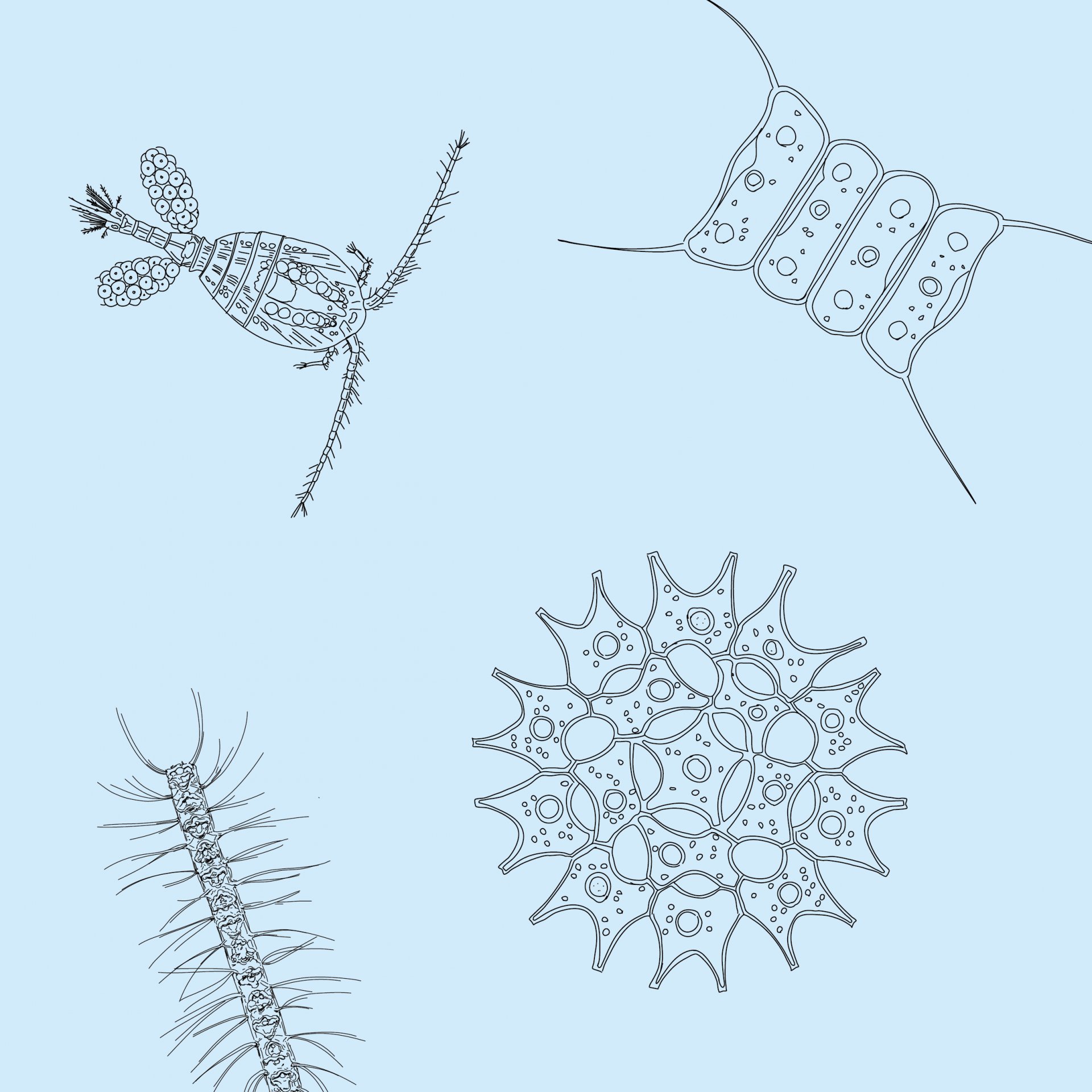
We are always looking for exciting knowledge about individual microbes, but also about entire ecosystems. The genetic material of microbes, their DNA, for example conceals a lot of information about their lifestyle and abilities. To analyze microbes in detail, to get to the bottom of their DNA, their enzymes, their biochemical capabilities, we use many different methods. Here we present some of the methods we use at our institute.
Analysis of marine sugars
Every bacterium, every microorganism has its very own enzymes – its own tools – in order to cut polysaccharides. We can use such enzymes to measure sugar molecules in the sea. The bacterial enzymes are responsible for the breaking down of sugar compounds. They break the large, complex polysaccharides into small, simple monosaccharide units. These simple sugars are easier to measure than polysaccharides. More...
Analysis of microbial metabolism
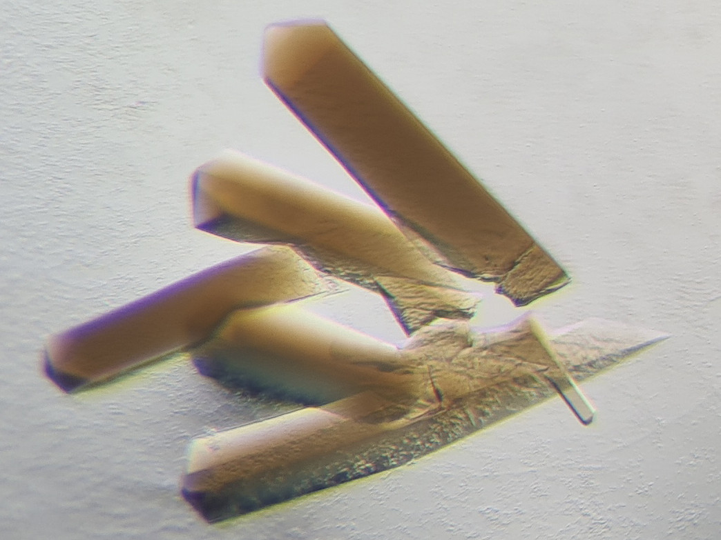
In focus are the enzymes involved in the conversion of minerals and gases. They are proteins that orchestrate strange reactions highly challenging for chemists. We have to extract these enzymes, and sort them out from other proteins by using the native purification. We then pierce the molecular secret of their reaction by looking at them with X-ray crystallography, which means that we have to crystallize the enzymes, and use X-ray to get their pictures. More...
Metagenomics
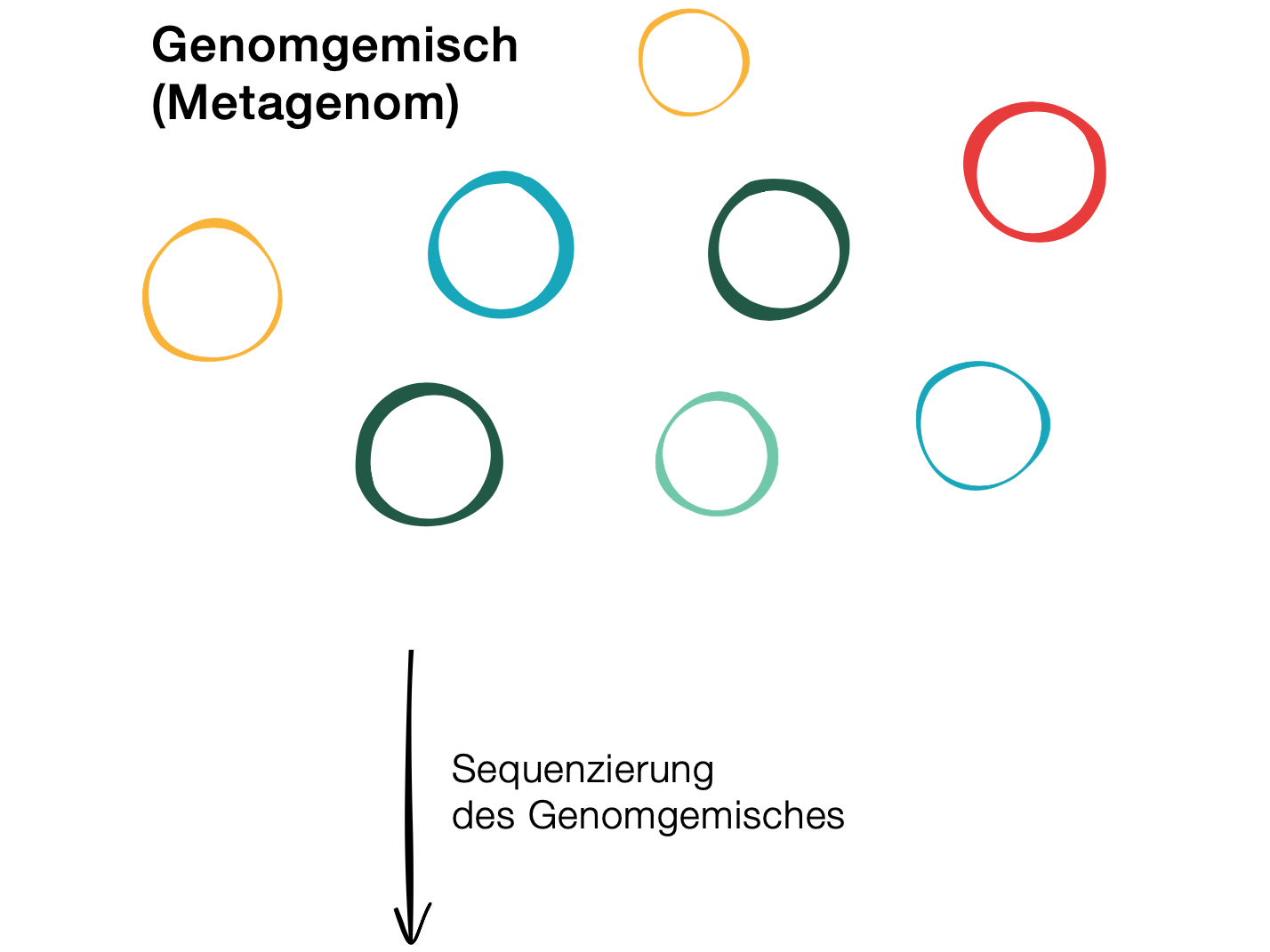
With the help of meta-genomics, it is possible to analyse an environmental sample (i.e. a mixture of the most diverse organisms as they live together in the environment) and find out which organisms live in this environment as well as which genes – and thus metabolic pathways, interactions, and defence strategies – predominate. More...
Fluorescence In Situ Hybridisation (FISH)
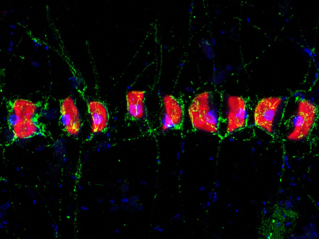
Microorganisms can barely be distinguished from each other based on their appearance. Nevertheless, every cell has its own fingerprint that is typical for the species – its genetic material. A case for FISH, Fluorescence In Situ Hybridisation. It selectively visualizes certain sections of the genetic material of individual cells. Under the microscope, these cells are then made to glow. FISH images of microorganisms look like a starry sky, but one with many different colours. Our question is: What is shining where? With FISH, we can precisely identify the organisms in our samples. More...
Cultivation and Isolation of Microorganisms
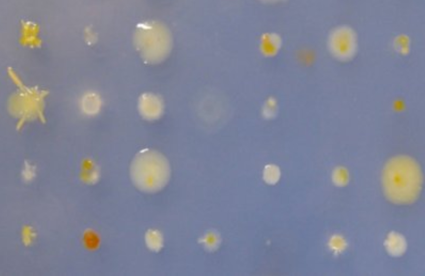
We dilute the cells with sterile seawater directly after sampling to isolate individual cells and incubate for long times to get slowly growing bacteria. The combination of physical isolation into hundreds of cultures, sufficient time to grow and molecular tools to analyze enrichment cultures still needs the perfect medium for the microorganism of interest. A perfect medium provides all nutrients at non-toxic concentrations, ideally in a permanent continuous-fed mode at micromolar concentration, like in nature. More...

