- Research & Instruments
- How we study - Our instruments and methods
- Instruments and Methods
- On Land
- Atomic Force Microscope (AFM)
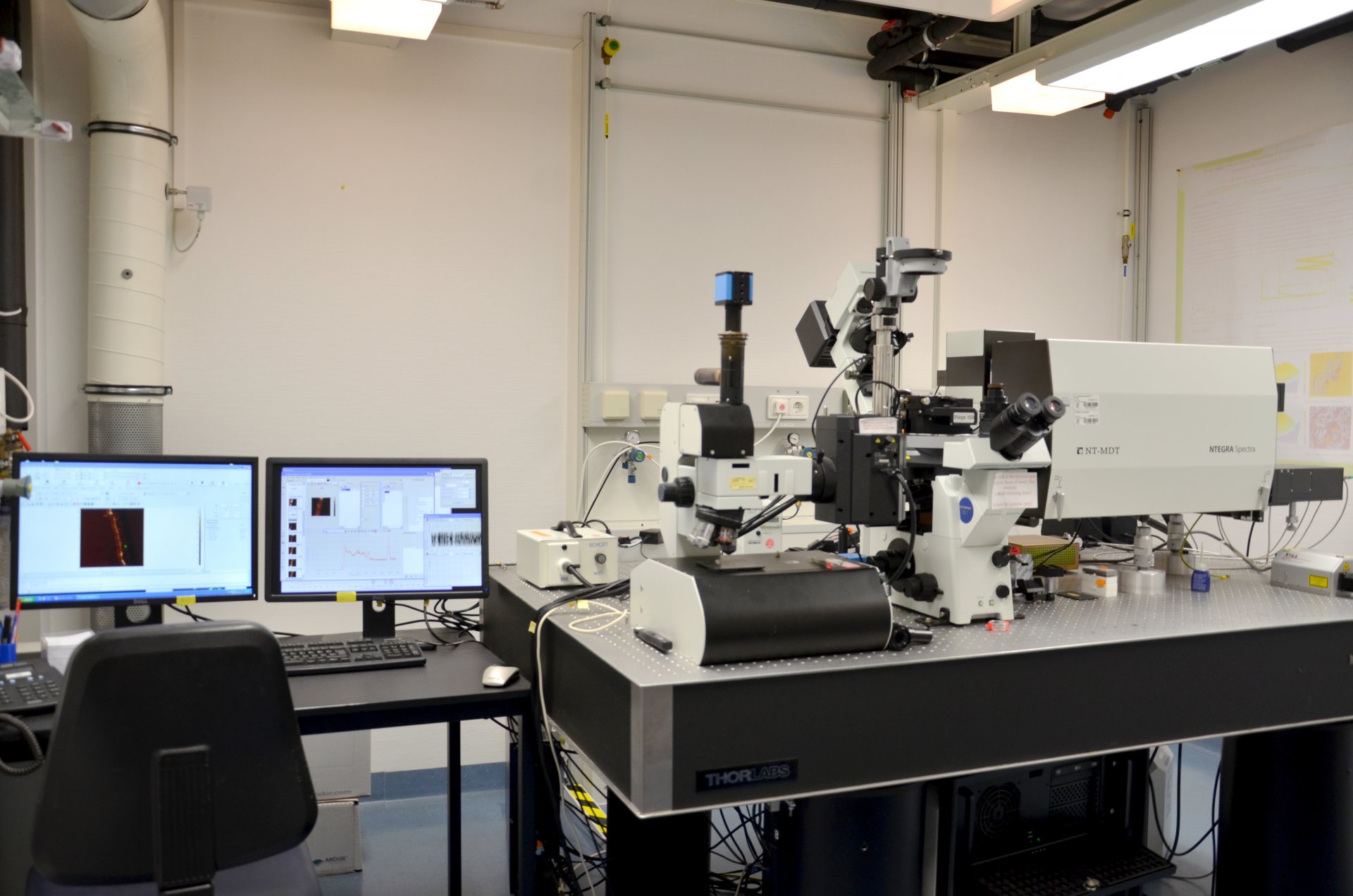
Atomic Force Microscope (AFM)
What is an atomic force microscope?
The atomic force microscope (AFM) is a scanning probe microscope. It was developed in 1985 by Gerd Binning, Calvin Quate, and Christoph Gerber.
Any solid material in air or liquids can be examined using atomic force microscopy. This is a great advantage for biological samples because they will not dry out and thus retain their shape. Information on the surface structure can be determined in the nanometre range.
In addition to the surface topography, physical properties such as the strength, elasticity, or magnetic strengths of a sample can be determined.
How does the atomic force microscope work?
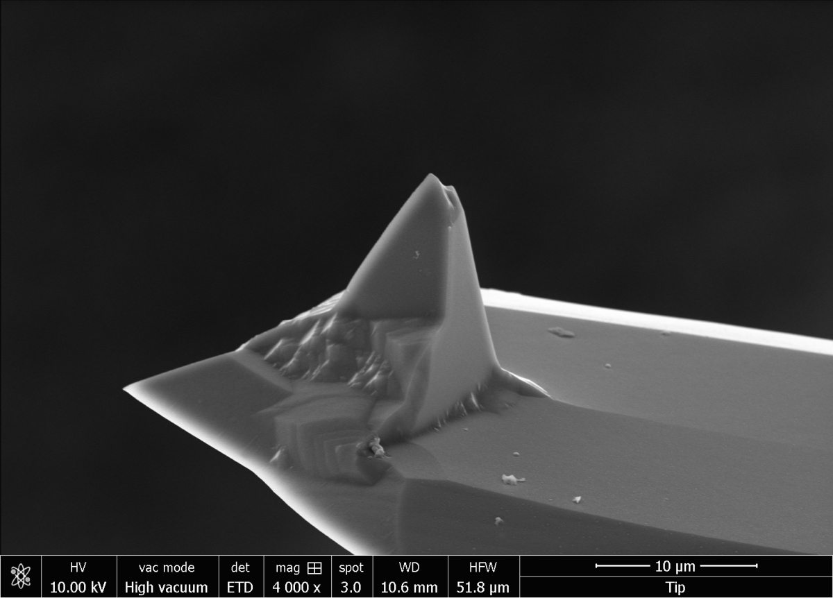
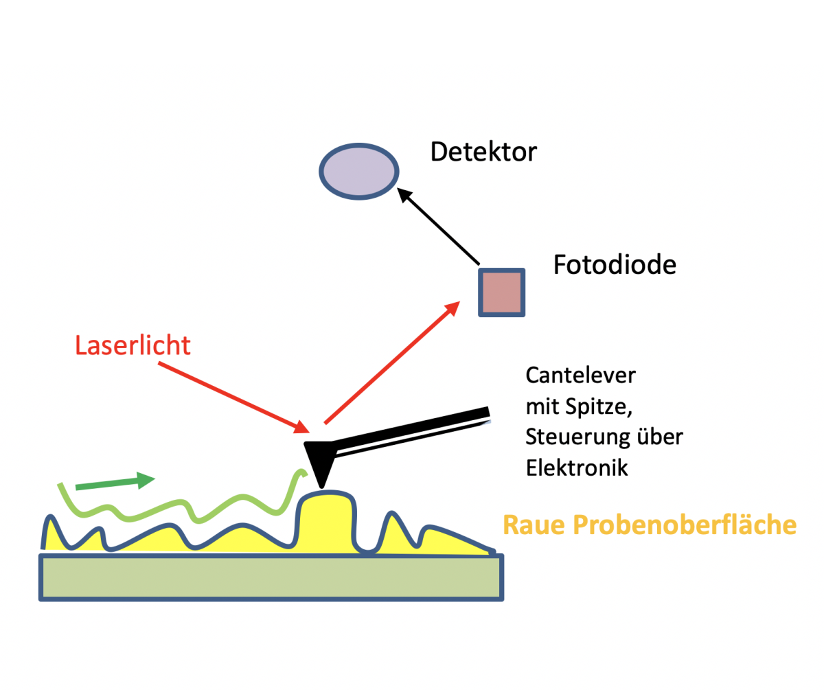
The measurement involves moving a spring (cantilever) with a very small needle attached to it over a sample surface. The bending of the spring is recorded by a laser and converted into a three-dimensional, microscopic image.
During scanning, the cantilever bends to varying degrees over the surface. These differences are a measure of atomic forces acting between the tip and the surface.
This movement is done point by point and line by line over a range of approx. 1 × 1 µm to 100 × 100 µm (scan). The spring is guided by piezo elements.
The atomic force microscope can be operated in different modes. This includes:
Contact mode
The measuring tip is in direct contact with the sample surface to be analysed. It can be regulated or unregulated.
In the controlled contact mode (constant force mode), the spring is steered by a piezo element so that the force between the tip and the sample remains constant. The change in position of the piezo element contains information of the surface structure of the sample.
The unregulated mode is the constant height mode, which is very suitable for smooth, hard surfaces. The forces of scanning the surface increase with increasing unevenness of the surface.
Intermittent mode (tapping mode)
This mode is mainly used for sensitive or unstable samples such as biological samples. Samples can also be examined in a liquid. In the intermittent or tapping mode, the excitation is set outside at a fixed frequency close to the resonant frequency of the spring. The resonance frequency of the spring between the tip of the spring and the sample surface to be examined changes because of interaction forces.
In this mode, the sample is not touched. It is therefore a gentle method for sensitive samples. Many scans with the same spring are possible.
The atomic force microscope in action
The following images show various bacteria scanned with the AFM. The resolution is quite high, and you can easily see the surface structure and differences.
This is where the focus of the measurements lies.
Through an interface of the HD mode, it is also possible to obtain additional information for each individual point measurement during the scan.
One of these is, for example, Young’s Modulus, the elasticity modulus, which can provide information about the strength of the bacteria.
Who uses the atomic force microscope?
Scientists, PhD students, technicians from the Department of Biogeochemistry, MarMic students during the lab rotation, and visiting students.
Contact
Group Leader
Greenhouse Gases Research Group
MPI für Marine Mikrobiologie
Celsiusstr. 1
D-28359 Bremen
Deutschland
|
Room: |
3128 |
|
Phone: |

Technician
MPI for Marine Microbiology
Celsiusstr. 1
D-28359 Bremen
Germany
|
Room: |
3131 |
|
Phone: |
