- Abteilungen
- Abteilung Molekulare Ökologie
- Molekulare Ökologie
- Hochauflösende Mikroskopie
Hochauflösende Mikroskopie
Mithilfe der hochauflösenden Mikroskopie ist es uns möglich kleinste Strukturen unterhalb der optischen Auflösungsgrenze sichtbar zu machen.
Die Techniken hochauflösende strukturierte Beleuchtung (SR-SIM) und photoaktivierte Lokalisationsmikroskopie (PALM) sind mit unserem konfokalem Laser Scanning Mikroskop kombiniert. mehr...
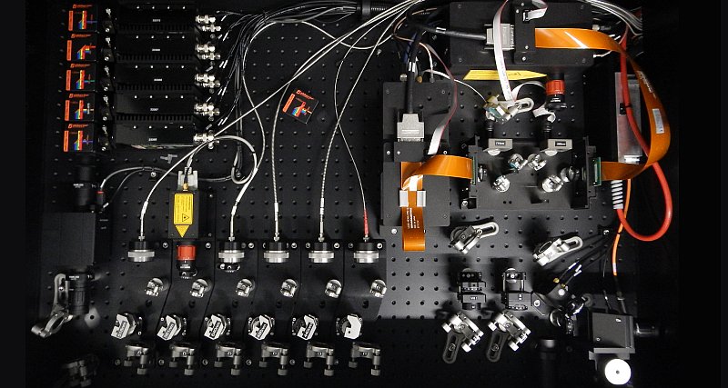
Unser neuestes Instrument basiert auf der Technik: Stimulated Emission Depletion (STED)[1].
Mit dem System easy3D STED der Firma Abberior Instruments kann eine lateral Auflösung unterhalb von 25 nm und eine 3D-Auflösung von bis zu 60 nm erreicht werden.
Das Gerät beherrscht die Methoden pulsed-STED[2], gated-STED[3],[4] und RESCue STED[5]. Es ist das erste kommerziell erhältliche STED-Mikroskop mit MINFIELD-Technik[6].


Technische Daten
| Anregungslaser | STED-Laser | Detektion* | |||
|---|---|---|---|---|---|
|
|
|
450/50 nm |
|||
|
|
(gepulst, 1 W) |
509/22 nm |
|||
|
|
525/50 nm oder |
||||
| 518 nm (gepulst, 300 µW) |
545/24 nm |
||||
|
|
(gepulst, 3 W) |
|
|||
| 615/20 nm | |||||
|
|
|
||||
*single-photon-counting avalanche photodiode (apd module)
Reservierung
Standort
Raum 2242, Tel. 931
Verantwortlich
Referenzen
| 1. | Hell, S.W., J. Wichmann. (1994). Breaking the diffraction resolution limit by stimulated emission: Stimulated-emission-depletion fluorescence microscopy. Optics Letters. 19: 780–82. (doi:10.1364/OL.19.000780). |
| 2. | Dyba, M., S. W. Hell. (2003). Photostability of a Fluorescent Marker Under Pulsed Excited-State Depletion through Stimulated Emission. Applied Optics. 42:5123–29. (doi:10.1364/AO.42.005123). |
| 3. | Vicidomini, G., G. Moneron, K.Y. Han, V. Westphal, H. Ta, M. Reuss, J. Engelhardt, C. Eggeling, and S.W. Hell. (2011). Sharper low-power STED nanoscopy by time gating. Nat. Meth. 8:571–3. (doi:10.1038/nmeth.1624). |
| 4. | Moffitt, J.R., C. Osseforth, and J. Michaelis. (2011). Time-gating improves the spatial resolution of STED microscopy. Opt. Express. 19:4242–54. (doi:10.1364/OE.19.004242). |
| 5. | Staudt, T., A. Engler, E. Rittweger, B. Harke, J. Engelhardt, S.W. Hell, (2011). Far-field optical nanoscopy with reduced number of state transition cycles. Opt. Express. 19:5644–57. (doi:10.1364/OE.19.005644). |
| 6. | Göttfert, F., T. Pleiner, J. Heine, V. Westphal, D. Görlich, S.J. Sahl, S.W. Hell. (2017). Strong signal increase in STED fluorescence microscopy by imaging regions of subdiffraction extent. Proc. Natl. Acad. Sci. USA. 114:2125-30. (doi:10.1073/pnas.1621495114). |