Page path:
- Press Office
- Press releases 2009
- 31.03.2009 Advanced colour photography – a ne...
31.03.2009 Advanced colour photography – a new method of spectral analysis gets to the bottom of single cells and biofilms
Advanced colour photography – a new method of spectral analysis gets to the bottom of single cells and biofilms
It requires little more than water and sunlight - and soon most surfaces will be covered in a thin, slimy coating. It's the green you see shimmering on pebbles in a stream, it's the brownish hue on the sand on the beach, or the dirty coat at the bottom of the bathtub. All of these are examples of biofilms – a thin layer of microorganisms.
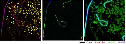
Studying how such biofilms develop, who inhabits them and how they interact with the environment is a tricky task. After all, they are razor-thin and very fragile. A new spectral imaging system, designed by scientists at the Max-Planck-Institute for Marine Microbiology in Bremen, Germany, now considerably enhances the ability to study organisms in such biofilms. „For the first time, the so-called MOSI-system (short for Modular Spectral Imaging) enables us to understand the relationship between the differently coloured regions in biofilms and the type of microorganisms that inhabit them. Furthermore, we can zoom in to a specific region of interest to broadly identify single cells that give the biofilm its colour (Fig. 1)“, developer Lubos Polerecky explains. Using a spectral image – similar to a normal colour picture, but with up to 200 colours, instead of just the usual three (see background below) – pigments in single cells can be quantified. Thereby, their identity and potential function can be asserted to some extent. The new device and some possible applications are now presented in the journal „Applied and Environmental Microbiology“.

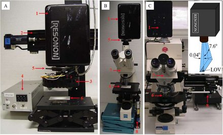
Another special feature of MOSI: It is handy and easily manageable and therefore easy to use in field studies. Packed in a medium-sized container, it can be reassembled and readjusted by a person within one to two hours. Moreover, MOSI is comparatively cheap, as it requires only a special hyperspectral camera, a moving stage and a lamp. For microscopic imaging, it can be mounted on most good quality microscopes that are typically available in any microbiology lab. Last but not least, the software required for fully automated measurements and advanced data analysis is provided for free by the MPI-scientists.
First applications
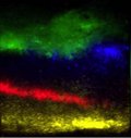
In their publication, Polerecky and his colleagues present a number of possible applications for the MOSI-system in the field of aquatic microbial ecology. The investigated objects can vary several orders of magnitude in size – ranging from single-cells up to the entire biofilm community – which gives scientists a chance not only to identify the different cell types but also understand how they interact with each other and with environmental factors such as light, oxygen, sulphide. For example, the scientists studied a microbial mat from the Spanish Lake Chiprana, the only natural permanently hypersaline (very salty) lake in Western Europe. Microbial mats are communities of cells that are typically structured in distinct layers of microorganisms: „phototrophic“ organisms, that build biomass from carbon dioxide using light energy, and „heterotrophic“ organisms, that degrade this biomass. Closed internal cycles of elements such as oxygen, carbon or sulphur make these mats ideal models for more complex, light-driven ecosystems. Spectral imaging clearly revealed the different layers of organisms, easily identifiable by their contained pigments (Fig. 2). The results indicated tight coupling between the different phototrophic groups in the mat, which is consistent with the results obtained by functional analyses.

Using sediment cores from Janssand, a sand bank close to the island of Spiekeroog in the German North Sea, the scientists combined spectral analysis with measurements of ecologically relevant parameters – namely the oxygen consumption rates. The MOSI-measurements supported the existing assumption that oxygen consumption is tightly coupled to algal abundance in suboxic sediment layers.

Summary
Existing methods allow detailed investigations of pigments only at the expense of the sample itself, which is destroyed during the analysis. Moreover, chemical pigment extraction procedures often require significantly larger sample volumes, more time and effort. MOSI provides a non-destructive identification, localization and relative quantification of pigments in microbial samples, thus allowing for successive investigations, such as when dealing with the temporal development of biofilms. Moreover, the procedure is not susceptible to pigment losses that may happen during chemical extraction. Fast, sensitive and non-destructive – MOSI opens new possibilities in studies investigating the role of spatial organization of microorganisms in the way complex microbial communities function and interact with the environment.
Background: Spectral Imaging
A spectral image shows the studied object in many more than just the usual 3 colours that we see when looking at a printed photograph (yellow, cyan and magenta) or while watching a TV (red, green and blue). The many different colours that are reflected, transmitted or emitted by an object are called a spectrum. By measuring and analyzing these spectra, much more can be learned about the identity and composition of the studied object. So far, this approach has been used for example in remote sensing of stars and planets, our Earth including, as well as in medicine and microbiology. In food technology, it helps the early identification of rotten meat, in the textile or furniture industry it unmasks additions of unwanted substances. The MOSI system, with its adjustable spatial resolution, can now also aid environmental microbiologists.
Fanni Aspetsberger
Fanni Aspetsberger
For further information please contact:
Dr. Lubos Polerecky 0421 2028 834
or the MPI press officers
Dr. Manfred Schlösser 0421 2028 704
Dr. Fanni Aspetsberger 0421 2028 704
Dr. Lubos Polerecky 0421 2028 834
or the MPI press officers
Dr. Manfred Schlösser 0421 2028 704
Dr. Fanni Aspetsberger 0421 2028 704
Original article:
Modular spectral imaging (MOSI) system for discrimination of pigments in cells and microbial communities. Lubos Polerecky, Andrew Bissett, Mohammad Al-Najjar, Paul Faerber, Harald Osmers, Peter A. Suci, Paul Stoodley and Dirk de Beer. Applied and Environmental Microbiology, Vol. 75, No. 3, p. 758-771, Feb. 2009.
Modular spectral imaging (MOSI) system for discrimination of pigments in cells and microbial communities. Lubos Polerecky, Andrew Bissett, Mohammad Al-Najjar, Paul Faerber, Harald Osmers, Peter A. Suci, Paul Stoodley and Dirk de Beer. Applied and Environmental Microbiology, Vol. 75, No. 3, p. 758-771, Feb. 2009.
Participating institutions:
Max Planck Institute for Marine Microbiology, Celsiusstrasse 1, 28359 Bremen, Germany
Center for Biofilm Engineering, 366 EPS - P.O. Box 173980, Montana State University-Bozeman, Bozeman, MT 59717-3980, USA
Center for Genomic Sciences, Allegheny-Singer Research Institute, 1107 11th Floor South Tower, 320 East North Avenue, Pittsburgh, PA, 15212-4772, USA
The press release as a pdf:
Original article:
Polerecky et al., AEM 2009
Polerecky et al., AEM 2009
Fig. 1: The shape and pigment content of single cells can be well resolved even in complex mixtures of cells – cyanobacteria in the given example. (red=chlorophyll a, green=phycocyanine, blue=phycoerythrine). (Source: L. Polerecky, AEM 2009)
Fig. 2: Hypersaline microbial mat from Lake Chiprana, Spain. Although the stratification is clearly visible to the naked eye (D, in original colour), spectral imaging allows more specific identification of these layers (E-G): the upper 3 mm are dominated by diatoms, 3-6 mm are inhabited mainly by cyanobacteria, the lower margin by Chloroflexus-like bacteria. (Source: L. Polerecky, AEM 2009)
Fig. 3: MOSI in action in mesoscopic (left) and microscopic (middle and right) investigations. (Source: L. Polerecky, AEM 2009)
